This site uses cookies to improve your experience. To help us insure we adhere to various privacy regulations, please select your country/region of residence. If you do not select a country, we will assume you are from the United States. Select your Cookie Settings or view our Privacy Policy and Terms of Use.
Cookie Settings
Cookies and similar technologies are used on this website for proper function of the website, for tracking performance analytics and for marketing purposes. We and some of our third-party providers may use cookie data for various purposes. Please review the cookie settings below and choose your preference.
Used for the proper function of the website
Used for monitoring website traffic and interactions
Cookie Settings
Cookies and similar technologies are used on this website for proper function of the website, for tracking performance analytics and for marketing purposes. We and some of our third-party providers may use cookie data for various purposes. Please review the cookie settings below and choose your preference.
Strictly Necessary: Used for the proper function of the website
Performance/Analytics: Used for monitoring website traffic and interactions
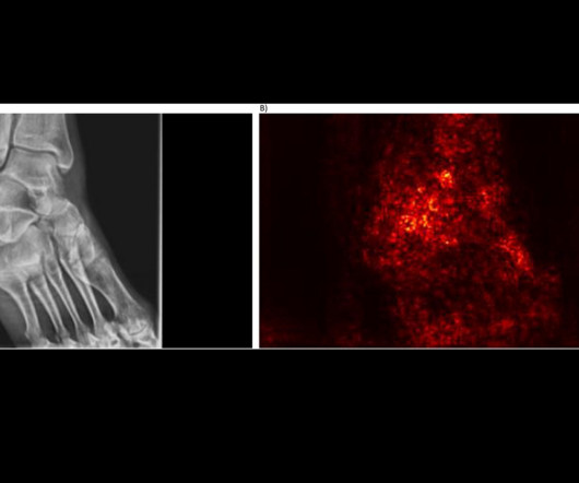
An AI model that detects low bone mineral density (BMD) on ankle and foot x-rays could be useful for screening for osteoporosis, according to radiologists at MD Anderson Cancer Center in Houston. First, the team culled a dataset from 907 patients over 50 years old who had undergone both DEXA scans and x-rays within 12 months.
A group in South Korea has validated a generative AI model that could reduce reading times and increase chest x-ray reporting accuracy, according to a study published March 11 in Radiology. To that end, Hong and colleagues, including researchers from Seoul-based companies Soombit.ai The full study is available here.
In a study described as a “competition between radiologists,” participants tasked with identifying abnormal findings on chest x-rays performed better with AI assistance than without AI assistance – though not by much and not in all cases, according to a group in Nanjing, Jiangsu, China.
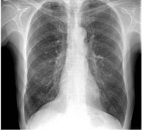
A commercially available AI algorithm shows potential for off-label use as a way to generate automatic reports for “unremarkable” chest x-rays, according to a study published August 20 in Radiology. These included chest radiographs that displayed abnormalities of no clinical significance, which are typically treated as normal.
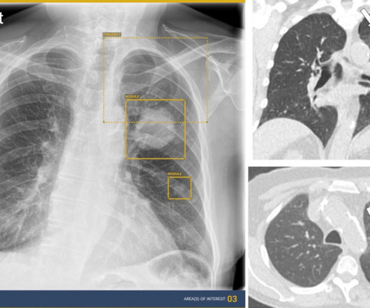
AI assistance can improve the detection accuracy of thoracic abnormalities on chest x-rays across radiologists with varying levels of expertise, according to a study published December 12 in Radiology. About 50% of the x-rays had abnormal findings. AI detected a lung mass, a lung nodule, and a small pneumothorax.
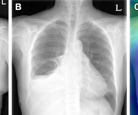
A group in Seoul, Korea, has developed an AI model that could help reduce radiology workflows by identifying “no changes” in follow-up chest x-rays of patients in critical care, according to a study published October 24 in Radiology. Example of triage of no change in a pair of chest radiographs in the emergency department.
Reading Time: 10 minutes read By Henry Williams, Carestream Area Vice President, Sales Western Nowadays, with hospital budgetary restrictions at the forefront of the purchasing decision making process, it seems like the X-Ray market, like everything else, is not immune to the current state of the economy. But is that really the case?
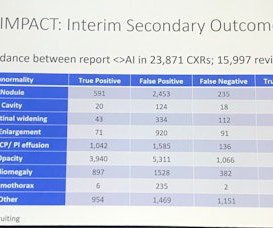
Nearly 72,000 chest x-rays had been randomized as of November 25 (the study is open through December 31), with the two primary outcomes of the trial being time to diagnosis of lung cancer and time from chest x-ray to CT by prioritizing abnormals. UCLH has produced 9,217 chest x-rays from 8,072 patients.
. | S1-SSCH01-5 | E451A This scientific paper may increase overall confidence in the potential of using multimodal AI for tuberculosis (TB) detection, and potentially autonomous reporting, on chest radiographs in certain clinical settings. respectively, where that of three radiologists' ranged between 91.9% to 94.7%, 89.4%
CHICAGO -- German researchers are testing ways to support nonradiologists in interpreting chest x-rays in emergency settings using an AI assistant. To test the AI algorithm, the LMU team focused on 563 cases, all ordered by the emergency unit department. Nonradiology residents 0.78
Four out of seven commercially available AI algorithms for detecting lung nodules on x-rays performed better than human readers, while two algorithms for predicting bone age fell short, in a study published January 9 in Radiology. Project AIR is an ongoing cohort study aimed at filling this gap, the authors wrote.
million chest radiographs. The team reported that the algorithm could successfully triage pairs of chest radiographs showing no change while detecting urgent interval changes during longitudinal follow-up. Julianna Czum, MD, from Johns Hopkins University wrote an editorial accompanying the study.
AI algorithms appear to have clinical value based on detecting normal x-rays – that is, by flagging chest x-rays as normal versus abnormal, they may reduce reading times for radiologists, according to research presented recently at the RSNA meeting in Chicago.
The first scans have been performed in the Olympic imaging polyclinic ahead of Friday's opening ceremony, and the 68-strong squad of radiologists and radiographers are primed and ready for action, according to musculoskeletal (MSK) expert Jérôme Renoux, MD. Bring it on!
AI sharply reduces radiologist reading times on chest CT Monday, November 27 | 1:30 p.m.-1:40 M6-SSNPM01-1 | Room E351 With help from an AI algorithm, radiologists can detect and classify lung nodules on routine clinical chest CT exams faster and more effectively, according to this new study. 1:40 p.m. | 1:50 p.m. | 3:50 p.m. |
Its impact on radiographer workflow ranges from detecting poor image quality on x-ray; automating CT imaging protocols; and for MRI, streamlining workflows for faster scan times, image reconstruction, and using synthetic MRI sequences.
Augmento X-Ray is designed to significantly reduce radiologist workload and improve the quality of chest X-ray reporting. billion annual X-rays performed, 1.5 The shortage of radiologists accentuates the significance of AI in tackling this challenge head-on.
S1-SSCH01-5 | E451A This scientific paper may increase overall confidence in the potential of using multimodal AI for tuberculosis (TB) detection, and potentially autonomous reporting, on chest radiographs in certain clinical settings. will discuss the impact of AI triage on chest x-rays for the purpose of accelerating lung cancer diagnosis.
In an open forum for radiographers at ECR 2024, Yi Xiang Tay, of Singapore University Hospital's radiography and diagnostic imaging department, shared his team's systematic review of the impact of imaging referral guidelines on patients and radiology services.
. | R1-SSCH09-5 | Room E352 An AI algorithm can find radiographic markers for osteoporosis that are common but often not reported on radiology reports, according to this scientific paper. The dataset consisted of 519 chest x-rays from patients ages 65 and older, collected from outpatient clinics.
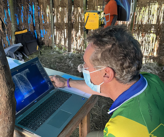
milla1cf Mon, 04/01/2024 - 11:44 April 1, 2024 — MinXray , a leading manufacturer of imaging systems for medical and veterinary use, recently sent its Impact Wireless X-ray system with a group of researchers and medical personnel to the YUS Conservation Area in Papua New Guinea.
Dubai-based Zscale Labs is launching Neuromorphic AI, an AI software application for multilabel classification of chest radiographs. Neuromorphic AI leverages the firm's hyperdimensional computing and deep-learning technologies to assist radiologists and healthcare providers in diagnosing multiple chest conditions from x-ray images.
The nurse is very qualified and skilled." In Sweden, an MR-sköterska must obtain a degree called röntgensköterska, so the references in the local media to an "x-ray nurse" being injured understates the formal competence of the man, who has not yet been named. "The accident was not caused by lack of knowledge.
It uses AI to analyze CT scans, X-rays and pathology slides, supporting clinicians in detecting and diagnosing medical conditions faster and more accurately. Radiologists using Harrison.ai's technology have seen an over 45% increase in diagnostic accuracy1. Averaged across all findings on chest radiographs.) [2]AIDE
Spirometry values were below predicted values and a standard chest radiograph depicted an elevated right hemidiaphragm that was not present on a prior CT examination during his SARS-CoV-2 infection. With the dynamic functional imaging capability of DDR, the authors could visualize thoracic and pulmonary motion and track diaphragm movement.
X-rays are the most widely used diagnostic tests, accounting for 60% of all imaging studies conducted. Radiologists and X-ray technologists are required to manage increasingly demanding caseloads while facing challenges from long hours and repetitive heavy lifting.
Teleradiology-India Introduction: “X-ray Visionaries” takes you on a compelling journey to unveil the expertise of radiographers and technologists, the unsung heroes of X-ray technology. The significance of their expertise in ensuring accurate diagnoses and patient well-being.
The studies include two oral presentations and five posters, and address AI advancements for chest x-ray reporting and breast cancer risk assessment, the company said.
A team of 32 radiologists and 36 radiographers are limbering up to work at the summer Olympics, which begins July 25. The imaging department at the polyclinic will be equipped with two mobile MRI scanners (Ingenia, from Philips), three ultrasound machines (Aplio i800, from Canon), and one x-ray system (from Primax International).
The other role could be that of a limited x-ray machine operator. It is ultimately going to be important for all of us to have a local geographic perspective as we think about these recruiting principles," Rubin said, adding that there is a "rising crisis" in terms of the volatility of the radiologist workforce. "We
MRI complements CT, ultrasound, and x-ray when it comes to trauma imaging," Mandel said. As undertrained radiographers represent an increased safety risk, a minimum of two MRI-trained radiographers must staff the emergency department MRI suite at all times. The bottom line? MRI is inherently unsafe," she said. "[For
A fundamental goal of radiographers is to complete an imaging exam that provides sufficient information for an accurate clinical diagnosis–and at the lowest possible dose. To make it easier for readers, I’ve organized the available solutions into three exam types: general X-ray, chest imaging, and pediatric imaging.
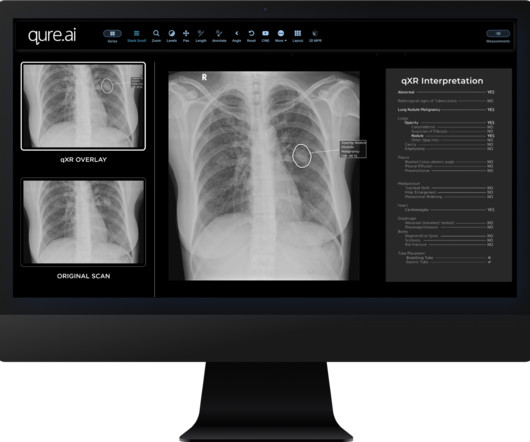
Food and Drug Administration (FDA) for its AI-enabled chest X-ray solution — qXR, under two critical findings. Digumarthy, MD , a senior co-author of the study and thoracic radiologist at Massachusetts General Hospital (Boston, MA), reported up to 96% sensitivity and 100% specificity for the qXR algorithm.
The authors note that ionizing radiation is the basis for the production of diagnostic X-rays, however it has long been proven to increase the risk of cancer. 26 –29, 2023 in North Hall, Level 3, booth #7913.
X-ray imaging helps clinicians locate and confirm osteomyelitis and assess its scope; MRI with contrast is used to confirm x-ray results. But a consistent interpretation system for MRI results has been lacking. Interreader agreement for using the MSKI-RADS system among the 20 readers was moderate, with an ICC of 0.7,
The Reveal Mobi Pro integrates KA Imaging’s Reveal 35C detector with SpectralDR technology into a complete mobile X-ray solution. The Reveal 35C detector mimics the workflow, dose, and techniques of state-of-the-art mobile DR X-ray. Our radiographic spectral images separate materials such as water (i.e.,
Shamie Kumar describes how AI fits into a radiology clinical workflow and her perspective on how a clinical radiographer could use this to learn from and enhance their skills. If the AI findings are seen in PACS, how many radiographers actually log into PACS after taking a scan or X-ray? Can Radiographers Up-Skill?
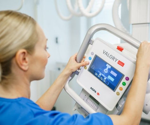
Agfa’s comprehensive portfolio supports “The Next Generation” in medical imaging by using intelligent and innovative technologies to ensure every X-ray image counts. Our productivity features and ‘one image is all it takes’ approach empower each X-ray expert to work more efficiently. A true One Click Workflow!
Closeup of X-ray photography of human brain Introduction: In the world of modern medicine, there exists a fascinating blend of art and science, where the careful use of technology and technique converges to reveal the hidden truths within the human body. Radiographic film, once the primary medium, has given way to digital sensors.
We organize all of the trending information in your field so you don't have to. Join 5,000 users and stay up to date on the latest articles your peers are reading.
You know about us, now we want to get to know you!
Let's personalize your content
Let's get even more personalized
We recognize your account from another site in our network, please click 'Send Email' below to continue with verifying your account and setting a password.


Let's personalize your content