This site uses cookies to improve your experience. To help us insure we adhere to various privacy regulations, please select your country/region of residence. If you do not select a country, we will assume you are from the United States. Select your Cookie Settings or view our Privacy Policy and Terms of Use.
Cookie Settings
Cookies and similar technologies are used on this website for proper function of the website, for tracking performance analytics and for marketing purposes. We and some of our third-party providers may use cookie data for various purposes. Please review the cookie settings below and choose your preference.
Used for the proper function of the website
Used for monitoring website traffic and interactions
Cookie Settings
Cookies and similar technologies are used on this website for proper function of the website, for tracking performance analytics and for marketing purposes. We and some of our third-party providers may use cookie data for various purposes. Please review the cookie settings below and choose your preference.
Strictly Necessary: Used for the proper function of the website
Performance/Analytics: Used for monitoring website traffic and interactions
Some of my radiological heroes would report a staggering 30,000 to 40,000 radiographs a year. So as to report more CT and MRI, radiologists stopped doing hands-on ultrasound and fluoroscopy. Some even [startled gasp] gave up reporting plain radiographs. I still don’t know how they did it. The neocortex rarely had to engage.
A team led by Junqi Han, MD, from the Affiliated Hospital of Qingdao University in China found that its model combining data from mammography images, ultrasound images, and other characteristics performed well in predicting disease-free survival of breast cancer. This means the combined model could predict prognosis after surgery.
Ultrasound model predicts liver disease progression. Appropriateness and imaging outcomes of ultrasound, CT, and MR in the emergency department: a retrospective analysis from an urban academic center. Commercially Available Chest Radiograph AI Tools for Detecting Airspace Disease, Pneumothorax, and Pleural Effusion.
Spirometry values were below predicted values and a standard chest radiograph depicted an elevated right hemidiaphragm that was not present on a prior CT examination during his SARS-CoV-2 infection. With the dynamic functional imaging capability of DDR, the authors could visualize thoracic and pulmonary motion and track diaphragm movement.
Substituting less energy-consuming ultrasound for x-ray or CT reduced energy use by as much as 8% during diagnostic radiology processes and 31.2% during indirect radiology department activities, according to findings of a pilot study presented March 1 at ECR 2024. kilowatts per patient, Masperi said.
The team's review of MRI's role for this indication was published January 3 in RadioGraphics. MRI findings regarding the state of pelvic nerves involved in endometriosis are key to planning treatment, w rote a group led by Ceylan Colak, MD.
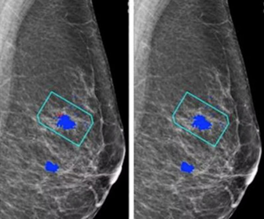
For the AI study, two views of each breast (four images) are taken to be evaluated independently either by the AI system plus a single reader or by a radiologist/radiographer as a first reader and second reader using the two-reader model. But even in nondense breasts, adding ultrasound improved the screening performance to nearly 78%."
She's also served as clinical director of CT, MRI, and ultrasound, and she was program director of radiologic technology and director of training for the Society for Imaging Informatics in Medicine (SIIM). Stewart earned her doctorate in biomedical (health) informatics from Rutgers University with a focus on clinical informatics.
Healthcare disparities continue to plague medical imaging, but there are concrete measures radiologists can take to mitigate them, according to a paper published on October 12 in RadioGraphics. B) A craniocaudal magnified mammogram more clearly shows the irregular mass and pleomorphic calcifications in the medial breast. (C)
The studies include two oral presentations and five posters, and address AI advancements for chest x-ray reporting and breast cancer risk assessment, the company said.
MRI complements CT, ultrasound, and x-ray when it comes to trauma imaging," Mandel said. As undertrained radiographers represent an increased safety risk, a minimum of two MRI-trained radiographers must staff the emergency department MRI suite at all times. MRI is inherently unsafe," she said. "[For
An ultrasound can take about 30 minutes to an hour while CT scans can take 10-20 minutes. This article will talk about the different diagnostic imaging methods such as X-rays, CT scans, Ultrasound, and MRI. X-ray Also called a radiograph, an X-ray uses radiation to create images of the body. It can take up to 90 minutes, max.
Positions (On-site): Body (100% Body) – Regions Hospital Mix of shifts worked on-site Mixture of hospital, outpatient, and remote Interpret MRI, CT, U/S, and radiographs After-hours coverage provided internally by the emergency radiology section No neuro or MSK Body/Mammo – Western Wisconsin 45-minute drive from the Twin Cities.
We are excited to support KA Imaging and their enhanced detector technology for the conventional and mobile radiographic markets,” said Marc Lorenzo, Executive Vice President at Del Medical. Before joining the company, Thompson was the President for Siemens Healthineers Ultrasound.
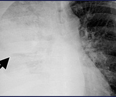
A chest radiograph (Fig 1a) showed a large, right pleural effusion (asterisk) and associated atelectasis. Ultrasound (Fig 1b) showed a simple pleural effusion (asterisk).
The product can be used to visualize known or suspected lesions of the breast in adults, as an adjunct to mammography and/or ultrasound. RadioGraphics 2019 39:7, 1907-1920. RadioGraphics 2021 41:3, 829-839. women older than 40 with dense breasts.2 Breast Density on a Mammogram. Updated April 4, 2023. Sensakovic, et al.
The Essentia SA is an ultra-compact straight arm system, designed for a wide range of standing, sitting and recumbent radiographic exams. Also at Fujifilm’s Booth #1929, the company will be celebrating 20 years of leadership in ultrasound elastography and will be conducting live scans with Fujifilm’s ARIETTA DeepInsight line.
Kim Mason Kim Mason, an Audit and Research Radiographer for Mid Yorkshire Teaching Hospitals Trust, talks about their role as well as the value of radiographer engagement in research activities and how to get involved. Hi, I’m Kim and I am an alternative-styled, funky-haired, septum-pierced, disabled Audit and Research Radiographer.
We recently adopted a nationwide pediatric appendix ultrasound performance protocol, sonographer worksheet, and radiologist reporting template in order to decrease CT utilization for this diagnosis nationwide. You can enroll in the free, on-demand course at Pediatric Appendix Ultrasound Standardized Performance and Reporting Training.
Although the word “radiology” sounds like it involves radiation, that is not always the case – for example, MRI (magnetic resonance imaging) and ultrasound do not use radiation in their medical imaging technologies. A radiographer is a medical professional who performs the scanning on patients.
Many healthcare professionals believe that radiologists sit in dark, dungeon-esque rooms, glaring at bright screens and reporting lists of radiographs in a monotonous fashion without any concern about a patient’s clinical situation. A common misconception is that radiologists do not have patient contact. That statement is false.
At Clermont Radiology’s Women’s Center, we offer Digital Mammography, Ultrasound, and DEXA Scans. Below is a description of each type of exam: Digital Mammography Mammography is the practice of radiographic imaging of the soft tissue of the breast. There is no injection or radiation exposure associated with ultrasound.
Photoprint from radiograph by W.K. 3) In the early twentieth century, it was a common goal for investigators to try to find a way to separate the superimposed shadows that were recorded when a complex structure was shown on a radiograph. (3) This was a defining publication in the field of medical ultrasound. (14) Röntgen, 1895.
A: Ultrasound showing deviation of the right lens margin posteriorly into the anechoic vitreous humor (red arrow). B: Ultrasound demonstrates the normal location of the left lens in the iris (green arrow). Lens subluxation can be diagnosed by ultrasound which shows deviation of the lens (Fig. Radiographics. 1, 2) [4].
Discuss their contributions to the development of radiographic techniques and their role in establishing women in the field. Ultrasound and Imaging Physics: Shaping the Science: Explore the contributions of women in the field of imaging physics. Discuss their impact on early breast cancer detection and improved patient outcomes.
Randomized, Controlled Trial of Ultrasound-Assisted Catheter-Directed Thrombolysis for Acute Intermediate-Risk Pulmonary Embolism. A prospective, Single-Arm Multicenter Trial of Ultrasound-Facilitated, Catheter-Directed, Low-Dose Fibrinolysis for Acute Massive and Submassive Pulmonary Embolism: The SEATTLE II Study. CHEST 2010.
Yes, there is an acute shortage of manpower in this space and if recent reports are anything to go by, the volume of radiographic procedures being conducted will surpass the number of radiologist being inducted into the field by a ratio of 15:2. The situation would have been easily averted with the usage of teleradiology services.
In these women, screening tests, such as ultrasound or MRI, when added to mammography, substantially increase the detection of early-stage breast cancer. A growing number of medical organisations link to the DenseBreast-info.org website, including the EFRS (European Federation of Radiographer Societies) and the Society of Radiographers.
R)(M)(ARRT), CRT(M), FSBI, FNCBC Back in the olden days, all patient communication had to go through the patient’s referring physician including biopsy recommendations, ultrasound referrals, additional views and technical repeats, which were almost always related to mammography positioning. Written by: Louise Miller, R.T.(R)(M)(ARRT),
Ultrasound has been shown to rapidly diagnose lens dislocation as it can visualize internal structures of the globe [2, 7]. Radiographics. The axial CT scan above demonstrates a right lens dislocation (Fig. The sagittal reformatted images show the right lens subluxed posteriorly and inferolaterally (Fig. Afr J Emerg Med.
Antenatal ultrasound may show one or more intracranial cysts that communicate with the ventricular system and/or subarachnoid space. Ultrasound and Magnetic Resonance in Prenatal Diagnosis of Congenital Anomalies.” Seeing the radiographic images made medical education come to life for him. Mikic, Aleksandra Novakov, et al.
They will also be needed for performing studies which require operator skill, such as ultrasound and IR procedures. 2011, Radiographics, pp. It is likely that they will lead clinic with patients to discuss their imaging, as already happens in some hospitals. Hough, Andrew. 2011, The Telegraph. Monsky, Derek S. Vien, Daniel P.
The first scans have been performed in the Olympic imaging polyclinic ahead of Friday's opening ceremony, and the 68-strong squad of radiologists and radiographers are primed and ready for action, according to musculoskeletal (MSK) expert Jérôme Renoux, MD. Bring it on!
This encompasses 120 million diagnostic X-rays, 91 million CT scans, 42 million MRIs, and 40 million ultrasounds -- numbers that continue to rise at a compound annual growth rate (CAGR) of roughly 4.2% (range 3%-5%, all imaging; 5%-10%, overnight/ER imaging). RadioGraphics. interpreted around 650 million imaging studies per year.
A team of 32 radiologists and 36 radiographers are limbering up to work at the summer Olympics, which begins July 25. The imaging department at the polyclinic will be equipped with two mobile MRI scanners (Ingenia, from Philips), three ultrasound machines (Aplio i800, from Canon), and one x-ray system (from Primax International).
Ultrasound rounds out the radiologist’s toolkit for supplemental imaging of women with dense breasts. Both handheld and automated ultrasound methods are shown to be effective in detecting mammographically occult cancer in women with dense breast tissue. The shortage of radiographers: A global crisis in healthcare. 2023.10.001.
Newborns' livers can be affected by a variety of congenital and acquired diseases, and imaging plays an important role in the workup and management of these, according to a study published November 7 in RadioGraphics. Ultrasound. Ultrasound is the primary modality for assessing newborns' livers. Contrast-enhanced ultrasound.
We organize all of the trending information in your field so you don't have to. Join 5,000 users and stay up to date on the latest articles your peers are reading.
You know about us, now we want to get to know you!
Let's personalize your content
Let's get even more personalized
We recognize your account from another site in our network, please click 'Send Email' below to continue with verifying your account and setting a password.


Let's personalize your content