This site uses cookies to improve your experience. To help us insure we adhere to various privacy regulations, please select your country/region of residence. If you do not select a country, we will assume you are from the United States. Select your Cookie Settings or view our Privacy Policy and Terms of Use.
Cookie Settings
Cookies and similar technologies are used on this website for proper function of the website, for tracking performance analytics and for marketing purposes. We and some of our third-party providers may use cookie data for various purposes. Please review the cookie settings below and choose your preference.
Used for the proper function of the website
Used for monitoring website traffic and interactions
Cookie Settings
Cookies and similar technologies are used on this website for proper function of the website, for tracking performance analytics and for marketing purposes. We and some of our third-party providers may use cookie data for various purposes. Please review the cookie settings below and choose your preference.
Strictly Necessary: Used for the proper function of the website
Performance/Analytics: Used for monitoring website traffic and interactions
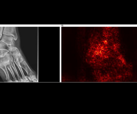
An AI model that detects low bone mineral density (BMD) on ankle and foot x-rays could be useful for screening for osteoporosis, according to radiologists at MD Anderson Cancer Center in Houston. First, the team culled a dataset from 907 patients over 50 years old who had undergone both DEXA scans and x-rays within 12 months.
A group in South Korea has validated a generative AI model that could reduce reading times and increase chest x-ray reporting accuracy, according to a study published March 11 in Radiology. Nonetheless, the era of generative AI in radiology holds great promise, they wrote.
Intelligent virtual and AI-based collimation features appear to save radiographers time during x-ray image acquisitions – a key function for enabling more patient-focused workflows, according to a recent study. ATC and SVO enable the radiographer to save time during chest or stitched examinations.
Plain hip x-rays may help screen patients for osteoporosis before they undergo total hip arthroplasty (THA), according to a study published November 14 in Scientific Reports. However, several studies have suggested that indices on plain hip x-rays may be reliable screening tools in female or Asian ethnicities.
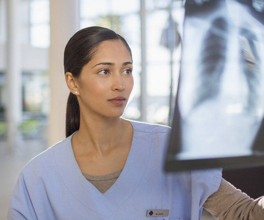
Reading Time: 4 minutes read New CDC Proposal Suggests Allowing Physician Assistants and Nurse Practitioners to Read Chest X-rays. This program trains and certifies physicians to read chest radiographs for workers participating in health surveillance programs. Would these professionals feel confident to interpret a chest x-ray?
A commercially available AI algorithm shows potential for off-label use as a way to generate automatic reports for “unremarkable” chest x-rays, according to a study published August 20 in Radiology. These included chest radiographs that displayed abnormalities of no clinical significance, which are typically treated as normal.
An AI model has delivered a long overdue update of pediatric bone growth predictions used in x-ray imaging to monitor scoliosis, according to a study published April 30 in Radiology. Full-body biplanar slot scanning is a type of low-dose digital x-ray imaging used to monitor scoliosis. Image courtesy of Radiology.
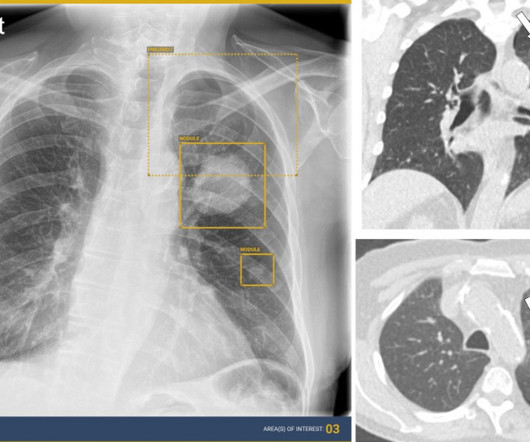
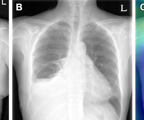
AI assistance can improve the detection accuracy of thoracic abnormalities on chest x-rays across radiologists with varying levels of expertise, according to a study published December 12 in Radiology. About 50% of the x-rays had abnormal findings. AI detected a lung mass, a lung nodule, and a small pneumothorax.
A group in Seoul, Korea, has developed an AI model that could help reduce radiology workflows by identifying “no changes” in follow-up chest x-rays of patients in critical care, according to a study published October 24 in Radiology. Example of triage of no change in a pair of chest radiographs in the emergency department. (A)
German developers of dark-field chest x-ray appear to have overcome a technical limitation of the technology – namely, adjusting for photon scattering caused by interferometers used in the experimental system. To overcome this limitation, the researchers developed a deconvolution-based correction method for the induced artifacts.
Nearly 72,000 chest x-rays had been randomized as of November 25 (the study is open through December 31), with the two primary outcomes of the trial being time to diagnosis of lung cancer and time from chest x-ray to CT by prioritizing abnormals. UCLH has produced 9,217 chest x-rays from 8,072 patients.
Reading Time: 10 minutes read By Henry Williams, Carestream Area Vice President, Sales Western Nowadays, with hospital budgetary restrictions at the forefront of the purchasing decision making process, it seems like the X-Ray market, like everything else, is not immune to the current state of the economy. But is that really the case?
In a study described as a “competition between radiologists,” participants tasked with identifying abnormal findings on chest x-rays performed better with AI assistance than without AI assistance – though not by much and not in all cases, according to a group in Nanjing, Jiangsu, China.
The agency's B Reader Program trains and certifies physicians to classify chest radiographs of workers participating in federal health surveillance programs.
CHICAGO -- German researchers are testing ways to support nonradiologists in interpreting chest x-rays in emergency settings using an AI assistant. Rudolph highlighted results for pneumothorax detection, in particular, where the AI assist performed similarly to the radiology resident readers. Nonradiology residents 0.78
The software could opportunistically screen patients at a smaller cost than other interventions, given it automatically operates "in the background" on radiographs performed for other reasons.
. | M4-SSMK03-4 | Room E450A A deep-learning AI model will be presented in this session that can predict bone mineral density T-scores from chest x-rays. They then trained the algorithm on a data set of 47,150 x-rays (23,151 patients) and validated it on an external data set of 2,914 radiographs (1,515 patients).
Current or past smoking does not appear to be significantly associated with radiographic progression in patients with psoriatic arthritis (PsA), according to a group of researchers from the University of Toronto. The study was published February 5 in Seminars in Arthritis and Rheumatism.
The college announced its opposition to the CDC's B Reader program exploring whether to let nurse practitioners and physician assistants read certain radiographs.
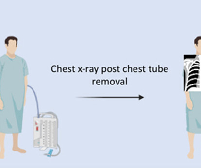
Ordering x-rays after removing chest tubes from lung surgery patients does not lead to meaningful change in patient management and prolongs hospital stays, according to a study published July 26 in the Journal of Thoracic and Cardiovascular Surgery. All patients who had any symptoms underwent a chest x-ray. days vs. 2.3
million chest radiographs. The team reported that the algorithm could successfully triage pairs of chest radiographs showing no change while detecting urgent interval changes during longitudinal follow-up. Julianna Czum, MD, from Johns Hopkins University wrote an editorial accompanying the study.
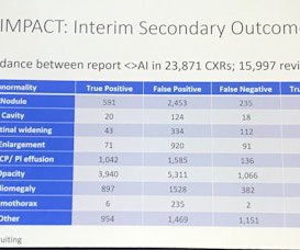
. | S1-SSCH01-5 | E451A This scientific paper may increase overall confidence in the potential of using multimodal AI for tuberculosis (TB) detection, and potentially autonomous reporting, on chest radiographs in certain clinical settings.
Carestream Health has launched a new x-ray system called DXR-Excel Plus with features designed to help simplify workflow and enhance productivity. Carestream's new DXR-Excel Plus x-ray system. Image courtesy of Carestream Health.
Four out of seven commercially available AI algorithms for detecting lung nodules on x-rays performed better than human readers, while two algorithms for predicting bone age fell short, in a study published January 9 in Radiology. Project AIR is an ongoing cohort study aimed at filling this gap, the authors wrote.
In an analysis of x-rays and bone scans, a group from Erasmus Medical Center in Rotterdam, the Netherlands, found that weight-bearing recreational activity was significantly associated with increased odds of knee osteoarthritis in individuals with low levels of lower-limb muscle mass.
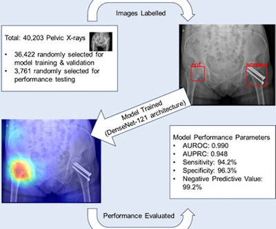
Fracture risk tool validated in men with prostate cancer Spine x-rays recommended in prostate cancer patients AI algorithm aids in classifying hip fractures on radiographs AI algorithm accurately detects trauma on pelvic radiographs
An X-ray collimator is an essential component of medical imaging equipment that directs and shapes X-ray beams to ensure precise targeting during radiographic procedures.
Food and Drug Administration (FDA) for its computer-aided detection chest x-ray qXR-LN software for identifying lung nodules. The software flags suspected pulmonary nodules ranging between six mm and 30 mm and can serve as a second reader for clinicians interpreting frontal chest radiographs.
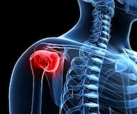
Researchers at the Mayo Clinic in Rochester, MN, have taken a first step in using AI to automatically classify and organize shoulder x-rays on a large scale, according to a recent study. on aTSA, and 100% on RSA x-rays. When classifying the imaging projection, the algorithm achieved F1 scores of 99.2%
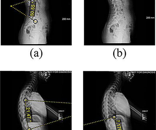
An AI model for x-ray imaging could help clinicians plan treatment other than spinal fusions in patients with adolescent idiopathic scoliosis, according to research published January 14 in PLOS One. For all patients, spinal x-rays were taken at six visits, from patients’ first standing x-ray to their most recent follow-up exam.
DDR is a novel functional imaging technique that uses sequential images obtained by a pulsed x-ray generator and a flat panel detector with a large field of view. frames per second (COPD patients) or 15 frames per second (healthy volunteers) with synchronized pulsed x-rays. Dynamic image data was acquired at 7.5
DDR is an emerging imaging technique that uses a pulsed x-ray source to acquire a series of radiographs at six to 15 frames per second. These images are then processed to visualize joints in motion.
The first scans have been performed in the Olympic imaging polyclinic ahead of Friday's opening ceremony, and the 68-strong squad of radiologists and radiographers are primed and ready for action, according to musculoskeletal (MSK) expert Jérôme Renoux, MD. Bring it on!
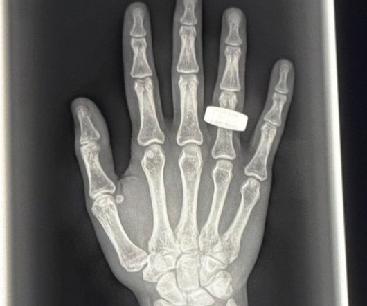
Food and Drug Administration (FDA) has cleared AZmed 's Rayvolve x-ray software for pediatric use. The research included a dataset of 3,000 pediatric radiographs and demonstrated Rayvolve's sensitivity of 96%, specificity of 86%, and area under the curve of 94%.
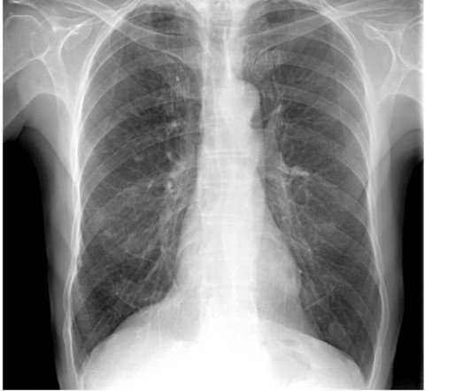
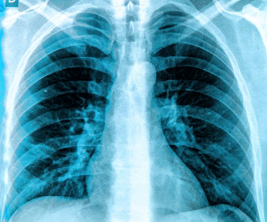
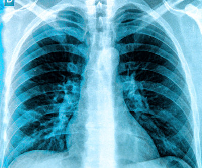
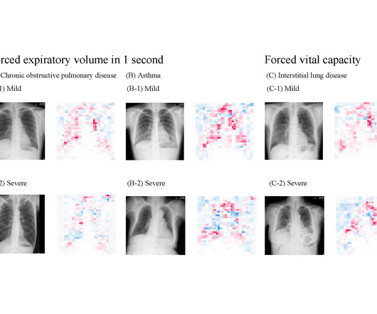
If there is one medical exam that everyone in the world has taken, it's a chest X-ray. Clinicians can use radiographs to tell if someone has tuberculosis, lung cancer, or other diseases, but they can't use them to tell if the lungs are functioning well.
Common diagnostic tests for pulmonary disorders include chest x-rays and pulmonary function tests (PFTs). a) Raw example of a dynamic digital radiograph. (b) The digital technology limits radiation exposure to patients compared with standard chest x-rays, they wrote.
AI algorithms appear to have clinical value based on detecting normal x-rays – that is, by flagging chest x-rays as normal versus abnormal, they may reduce reading times for radiologists, according to research presented recently at the RSNA meeting in Chicago.
M6-SSNPM01-2 | Room E351 In this talk, researchers will highlight the potential for AI software in detecting fractures that are often missed on radiographs. Opportunity for osteoporosis check on lumbar spine plain radiographs Wednesday, November 29 | 10:00 a.m.-10:10 1:50 p.m. | 3:50 p.m. | 10:10 a.m. | 10:10 a.m. | 10:20 a.m. |
Its impact on radiographer workflow ranges from detecting poor image quality on x-ray; automating CT imaging protocols; and for MRI, streamlining workflows for faster scan times, image reconstruction, and using synthetic MRI sequences.
We organize all of the trending information in your field so you don't have to. Join 5,000 users and stay up to date on the latest articles your peers are reading.
You know about us, now we want to get to know you!
Let's personalize your content
Let's get even more personalized
We recognize your account from another site in our network, please click 'Send Email' below to continue with verifying your account and setting a password.


Let's personalize your content